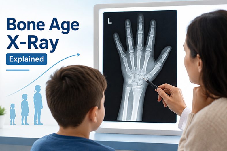
A bone age X-ray gets ordered, a radiologist writes back a single number, and suddenly a parent is holding a report that says their eight-year-old has the skeleton of a ten-year-old — with no explanation of what that actually means. This guide breaks down what the film is really measuring, how the two competing reading methods work, what a gap between bone age and chronological age does and does not tell you, and when this test is actually worth having.
What a Bone Age X-Ray Is Actually Measuring
Every bone starts as cartilage and gradually ossifies (hardens into bone) in a predictable sequence as a child grows, with the process finishing only once the growth plates fully fuse at the end of puberty. A bone age X-ray — almost always a single image of the left hand and wrist — captures a snapshot of exactly how far along that ossification sequence a child has progressed, compared to published reference standards built from large samples of healthy children at each age.
That is a meaningfully different thing than chronological age or even current height. A child can be tall for their age with a bone age right on target, or average height with a bone age noticeably ahead or behind — the X-ray is reading skeletal maturity specifically, which is what determines how much longitudinal growth potential is left in those growth plates.
Two Ways Radiologists Read the Same Film
| Method | How It Works | Practical Trade-off |
|---|---|---|
| Greulich-Pyle (GP) | Compares the whole hand image side by side against a published atlas of reference photos, matching to the closest overall pattern. Originally published in 1959. | Faster to perform, the most widely used method worldwide, but reliability varies somewhat by ethnicity since the original atlas was built from a specific population. |
| Tanner-Whitehouse (TW2/TW3) | Scores individual bones one at a time on a numeric maturity scale, then sums the scores into a bone age using conversion tables. | More granular and, in principle, more reproducible, but takes noticeably longer to score and requires more training to apply consistently. |
In a controlled study where the same films were read by both radiologists and pediatric endocrinologists, agreement between readers was high (an intraclass correlation of 0.95). But that same research began because clinicians had noticed real-world disagreements of more than a year between readers often enough to investigate it formally — a useful reminder that bone age involves genuine human judgment, not just an automatic measurement.
From Bone Age to a Height Estimate
Bone age itself is not a height prediction — it becomes one only when combined with the child’s current height in published conversion tables (most commonly the Bayley-Pinneau tables, first published in 1952), which estimate what percentage of adult height a child has already reached based on their bone age. We cover the accuracy of that prediction step, and how it stacks up against other height-prediction methods, in detail in our guide to How Accurate Are Height Prediction Calculators? — this article focuses specifically on the bone age reading itself.
What "Advanced," "Delayed," and "On Track" Actually Mean
A single film is a snapshot, not a diagnosis. Pediatric endocrinology literature is direct about this: bone age is genuinely useful for confirming diagnoses like constitutional delay of growth and puberty, interpreting hormone test results, and guiding decisions such as when to start treatment — but overemphasizing a single bone age reading, outside of that broader clinical picture, can be misleading. It is one input among several, not a standalone verdict.
Why a Doctor Actually Orders This
Bone age is not part of a routine well-child visit — it is ordered for a specific reason, usually one of the following:
Radiation, Cost, and What to Expect
The exam itself is quick and simple: a single X-ray image of the left hand and wrist, taken palm-down on a flat surface. Published estimates put the effective radiation dose at roughly 0.0001 to 0.1 mSv per exposure — for context, that is a small fraction of the natural background radiation a person receives over an ordinary day. It is not zero, which is part of why this test is reserved for children with an actual clinical question, rather than run routinely out of curiosity.
What This Means For You
The practical takeaway: a bone age reading is a genuinely useful clinical tool when it is ordered for a real reason and interpreted alongside a child’s full growth history — not a standalone verdict on how tall they will be or whether something is wrong. If a report comes back with a gap that surprises you, the right next step is a conversation with the ordering doctor, not a repeat X-ray or a second opinion calculator.
Frequently Asked Questions
What counts as a normal difference between bone age and chronological age?
Roughly a year in either direction is common and usually considered unremarkable. A gap of more than 2 years, advanced or delayed, is the general threshold pediatric guidance points to as worth a referral for further evaluation.
Does an advanced bone age always mean early puberty or a health problem?
No. Advanced bone age is associated with early or precocious puberty and, in some cases, with childhood obesity, but plenty of children run mildly ahead of their calendar age and go on to have entirely typical growth and health.
Can a bone age reading be wrong?
Reader judgment is part of the process, and studies comparing readers under controlled conditions still find room for disagreement, particularly between the Greulich-Pyle and Tanner-Whitehouse methods. This is one reason a single reading is treated as one input rather than a final answer.
How often is bone age repeated during treatment?
This varies by the reason it was ordered, but it is commonly repeated roughly once a year when it is being used to monitor puberty progression or a growth-related treatment, so the ordering clinician can track the trend rather than relying on one snapshot.
Is bone age the same thing as a growth plate scan?
Not exactly. A bone age X-ray reads the overall maturity of multiple bones in the hand and wrist as a proxy for skeletal maturity throughout the body; it is not a direct scan of the growth plates in the legs or spine, though it is used to estimate how much time those growth plates likely have left before they fuse.